
download
Simplify the Analysis of 3D Brain and Neuroimaging Data
How five neuroscience studies turned 3D data into reliable results
Modern imaging technologies produce incredibly rich, detailed, high-resolution datasets. With the right imaging workflows and analysis software, that detail becomes reliable insight.
See how five neuroscience studies used Amira™ Software to analyze neuroimaging data, including 3D brain images, and how similar approaches could simplify your own analysis.
Download the eBook to discover how:
- Nanoscale 3D analysis maps synaptic vesicles and scaffolding proteins in near-native synapses
- Volumetric reconstruction reveals endoplasmic reticulum remodeling and increased contact with the cell membrane after neuronal injury
- Nondestructive 3D tissue imaging captures microglial morphology in intact tissue
- 3D reconstruction workflows trace single neurons across Golgi-stained datasets and imaging modalities
- High-resolution episcopic microscopy data become quantitative 3D brain maps
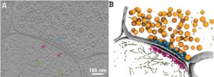
Ultrastructural analysis of synaptic tomograms after FIB milling. (A) Single XY slice from a reconstructed synapse tomogram. (B) Segmented rendering of the same tomogram. Gray shows plasma membranes; orange shows synaptic vesicles; cyan shows presynaptic protein density; magenta shows PSD protein density; green shows actin filaments. Figure reproduced from Held et al. under CC BY 4.0.