
download
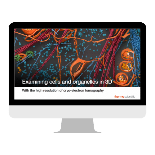
How to Visualize Molecular-scale Detail in 3D Within Intact Cells
Biomolecular detail revealed within the context of the cellular environment
Many aspects of cellular organization remain difficult to observe directly, particularly at the level where biomolecules, organelles, and cellular architecture come together.
Recent advances in cryo-electron tomography (cryo-ET) are helping to close this gap.
By preserving cells in a near-native, vitrified state and reconstructing them in three dimensions, cryo-ET reveals molecular-scale structural detail in situ, allowing you to visualize how macromolecular assemblies, membranes, and organelles are arranged and interact within the cell.
This ebook explores how these new imaging capabilities reveal biomolecular-level structural detail within cryopreserved cellular environments, adding a complementary layer of insight to existing biological imaging approaches.
In this guide, you’ll discover:
• How biomolecular assemblies can be visualized within the cellular environment
• Cellular architecture and organelles observed at sub-nanometer resolution in near-native, cryopreserved samples
• How molecular-scale structural detail can be placed in cellular context
• How insights from cryo-ET complement other imaging techniques
• How cryo-ET can be applied across cells, tissues, and organoid samples
Download the ebook to deepen your cryo-ET expertise and see cellular architecture in a new light.