download
See the Whole Picture: A 3D-to-Multiplex Spatial Biology Workflow for Precision Tissue Profiling
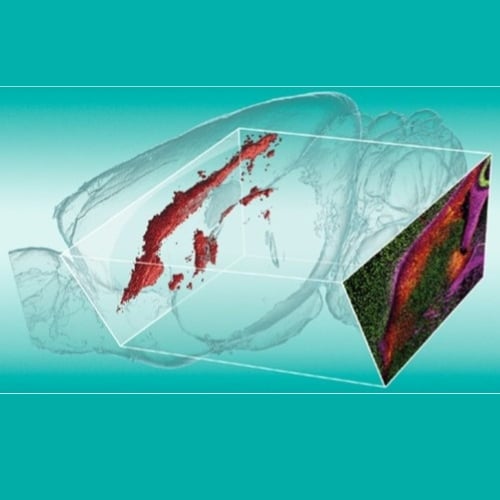
From whole-organ 3D imaging to single-cell resolution — without losing your place
Standard spatial biology starts with a thin section. But what if the biology you care about is somewhere else in the tissue entirely?
Download this application note and discover how to combine 3D light sheet imaging with high-plex spatial proteomics — on the same specimen — to profile tissue with unprecedented spatial precision.
In this application note, you will learn how to:
• Use whole-organ 3D imaging to map tissue architecture and pinpoint your region of interest before you section
• Guide anatomically precise cryosectioning using light sheet–derived spatial data
• Profile 100+ markers at single-cell resolution with MACSima® Imaging Cyclic Staining (MICS)
• Preserve epitope integrity and fluorochrome signal throughout the entire 3D-to-multiplex workflow